A team of materials scientists at Pennsylvania State University, led by Professor Venkatraman Gopalan, has developed a computational framework that interprets nonlinear optical microscopy images to determine the atomic structure and properties of materials. The research, demonstrates a method to extract quantitative information from microscopy images, providing insights into microscopic material behavior that were previously difficult and time-consuming to obtain.
Suceava, A., Hazra, S., Nag, J., Hayden, J., Imam, S., Liu, Z., Iyer, A., Kanatzidis, M. G., Trolier-McKinstry, S., Maria, J.-P., & Gopalan, V. (2025). Quantitative nonlinear optical polarimetry with high spatial resolution. Optica, 12(8), 1153. https://doi.org/10.1364/OPTICA.559060
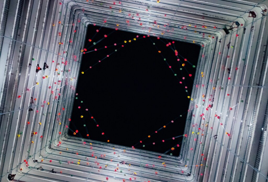
Nonlinear optical microscopy uses intense laser light to probe materials, producing signals that can reveal otherwise invisible structural features. Unlike conventional imaging, which relies on linear interactions such as reflection or absorption, nonlinear microscopy detects signals created when intense light interacts with atoms and molecules, producing new optical frequencies. These interactions can reveal material polarity, atomic vibrations, and other critical structural information.
“Nonlinear optical microscopy is an important tool that can reveal structural information about different materials,” said Albert Suceava, a doctoral student in materials science and engineering at Penn State and lead author of the study. “Our method allows us to interpret exotic interactions between light and matter, revealing details that were previously hidden. It can be applied to a wide range of samples, from biological materials to quantum computing devices.”
The project began when the team observed unexpected phenomena in microscopy images of well-understood samples. “We were seeing features that looked almost like optical illusions,” Suceava explained. “We needed to ensure that these signals were accurate and not artifacts of the microscope. Our framework models the effects of tightly focused laser light on polarization and interactions with the sample, allowing us to distinguish real material properties from optical effects.”
The framework is designed to translate the complex signals generated in second harmonic generation microscopy into quantitative material information. Second harmonic generation occurs when a material doubles the frequency of incoming light, effectively converting infrared into visible wavelengths. This process can reveal asymmetries in electron distribution, offering insight into material polarity and local atomic arrangements.
Professor Venkatraman Gopalan from Pennsylvania State University stated,
“Light is central to how we perceive and analyze the physical world. From electrons to atomic clusters, each component interacts with light at different frequencies. By studying these interactions, we gain a signature of the material’s structure and dynamic behavior.”
The researchers tested the framework on a variety of reference materials, comparing the extracted data to known properties. This process allowed them to validate the approach and ensure accurate quantitative interpretation. Suceava noted that their framework moves beyond simple image generation. “We are not just taking pictures; we are analyzing the signals to understand what the atoms are doing. By mapping material properties rather than simply capturing images, we can create a library of material behaviors that can be applied across multiple fields.”
The framework also addresses challenges in reproducibility and standardization. Nonlinear optical microscopy generates complex data that can be difficult to interpret consistently. By providing a structured computational approach, the team aims to improve the reliability of material characterization, enabling other researchers to extract quantitative information efficiently.
This development could have significant implications for materials research, offering new ways to analyze microscopic structures in electronics, photonics, and biological systems. Accurate modeling of light-matter interactions at the nanoscale can inform the design of advanced materials, improve quality control, and accelerate discoveries in multiple scientific disciplines.
“Our goal was to simplify the interpretation of nonlinear optical signals while maintaining accuracy,” Suceava said. “We believe this framework will help the community better understand and standardize the analysis of these images, ultimately supporting the development of new materials and technologies.”
By combining advanced computational modeling with nonlinear optical microscopy, the Penn State team has provided a tool that transforms complex optical signals into actionable scientific information, enhancing our ability to explore and understand the microscopic world. This approach represents a significant step forward in the characterization of material properties and in the broader field of optical imaging research.

Adrian graduated with a Masters Degree (1st Class Honours) in Chemical Engineering from Chester University along with Harris. His master’s research aimed to develop a standardadised clean water oxygenation transfer procedure to test bubble diffusers that are currently used in the wastewater industry commercial market. He has also undergone placments in both US and China primarely focused within the R&D department and is an associate member of the Institute of Chemical Engineers (IChemE).